


Using Ultrasound to Diagnose SI Joint Disease in Horses

AdobeStock image
By Kentucky Equine Research Staff
Transrectal ultrasound can reveal pathologic changes in the sacroiliac (SI) joint, including problems that may be present in horses that owners perceive as sound. Although ultrasound only images the caudal portion of the SI joint, or the section nearest the tail, one study found that changes detected in this region were representative of structural changes throughout the entire joint.* Based on these findings, the researchers concluded that transrectal ultrasound is a valuable first-line diagnostic tool for assessing SI pain.
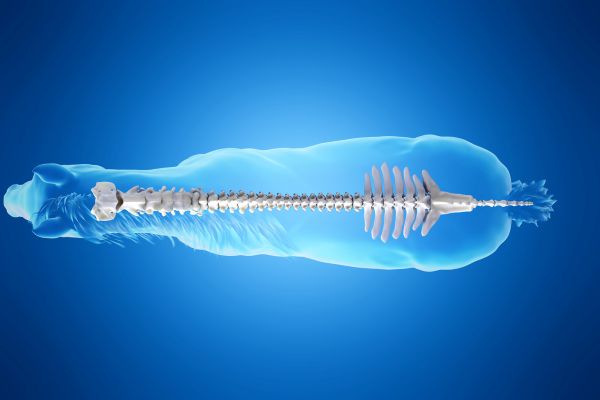
The SI joint is the articulation between the transverse processes of the first and second sacral vertebrae and the wings of the ileum. The sacral vertebrae are located at the base of the spine within the pelvis, just before the bones of the tail. Horses have two SI joints, one on the right and left side of the pelvis. Like any joint, horses can suffer injury and pain at this point, resulting in poor performance.
“Diagnosing SI pain can be challenging for veterinarians, as lameness from the SI joint can appear similar to thoracolumbar or lumbosacral pain, mild hind limb lameness due to proximal suspensory ligament inflammation, or even mild ataxia. Further, the joint is deep in the body, making it challenging to image,” said Kathleen Crandell, Ph.D., a nutritionist for Kentucky Equine Research.
During any musculoskeletal disease workup, ultrasound is a powerful diagnostic tool. In the case of the SI joint, however, transrectal ultrasound can only reach the caudal third of the SI joint. In a study by Mathys and colleagues, 25 SI joints from 15 Warmblood horses with no owner-reported lameness were examined by both computed tomography and ultrasound. Abnormalities were observed in 100% of SI joints using computed tomography and 92% of SI joints using ultrasound. Changes included osteophytes (abnormal bone growth in a joint secondary to osteoarthritis), joint effusion (fluid accumulation), sclerosis, subchondral bone lesions, and evidence of bone remodeling. Computed tomography images included the entire joint, and researchers found that the most severe changes occurred caudally, with only about one-quarter of the changes occurring cranially (the part of the SI joint closest to the head).
“A significant correlation between computed tomography findings in the caudal aspect of the joint and the entire joint was noted, meaning that the pathologic changes observed in the caudal one-third represent the changes in the entire joint in almost half of all horses. Examining the caudal one-third of the joint reachable by transrectal ultrasound, a much more economical and widely available diagnostic tool than computed tomography, can be used as a first-line tool to assess the SI joint,” explained Crandell.
As noted by the researchers, the abnormalities reflect joint remodeling, which means that strategies used to optimize joint health in other areas of the body, such as the stifles, hocks, knees, and ankles, also apply to the SI joint. Consider prophylactically administering oral joint health supplements to horses before signs of disease manifest. Kentucky Equine Research offers research-proven joint supplements containing a combination of glucosamine, chondroitin sulfate, and hyaluronic acid.
“This is important because the owners of the horses included in this study did not report any sign of lameness, yet almost all the horses had abnormalities of the SI joint diagnosed by computed tomography and/or ultrasound in this study. SI joint disease is highly prevalent and, while it may be part of normal aging or adaptation to training, taking steps to support these joints may allay performance issues,” advised Crandell.
*Mathys, R.A., T.R. Schmitz, H. Geyer, N. Borel, M. Hilbe, S. Ohlerth, and A.S. Bischofberger. 2023. Multi-detector helical computed tomography, transrectal ultrasonography, and histology of the sacroiliac joint: A comparative study in adult Warmblood horse cadavers. Animals (Basel) 15(13):1995.