


Understanding Navicular and Heel Pain in Horses

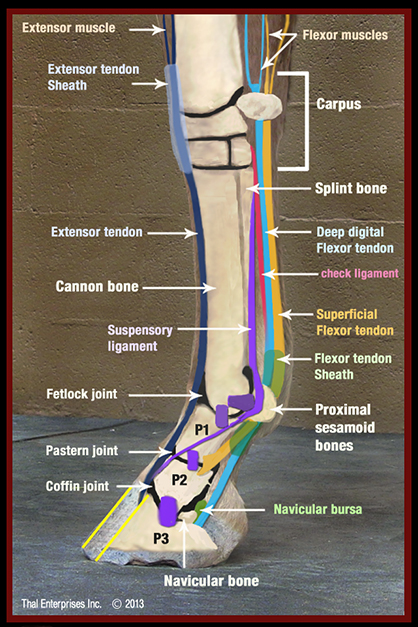
The major bone and soft tissue structures of the equine lower limb. Image courtesy HorseSideVetGuide.com.
By: Horse Side Vet Guide (edited)
No Hoof. No Horse. Maybe you have heard that old horseman’s saying. There is a lot of truth to it. The foundation of every horse is the foot.
Dr. Doug Thal, the creator of Horse Side Vet Guide, recently wrote a very comprehensive and well-illustrated article on Navicular Syndrome and Heel Lameness in Horses.
Here is an excerpt:
In order to understand navicular syndrome, it’s necessary to understand the basic anatomy of the foot. The equine “foot” includes the hoof capsule and its associated structures, the lower end of the short pastern bone, the coffin bone (P3, Pedal bone) and the navicular bone. There is the “hoof complex” itself – the hoof capsule, sole, frog, digital cushion, and ungual (collateral) cartilages.
The coffin bone takes up approximately 2/3 of the hoof, and it is approximately the shape of the hoof. Running down the back of the limb, diving into the heel of the foot and attaching to the “palm” of the coffin bone, is a wide, flat very dense tendon called the deep digital flexor tendon (DDFT= turquoise on the image to the right). The action of the DDFT is to flex the coffin joint and provide the floor of the joint. It courses over the navicular bone, using it as a fulcrum near its attachment to the coffin bone.
WHAT YOU MIGHT OBSERVE – SIGNS OF NAVICULAR DISEASE
“My horse just seems stiff, or short. He’s not really lame, he’s just off…”
Navicular syndrome usually first appears as a very mild lameness that gradually worsens over weeks to months. It usually affects both front feet, but one often seems worse than the other. As with most lameness, the lameness from navicular syndrome is most noticeable at the trot, resulting in a head-bob. Affected horses often have a short, choppy trot and canter, and tend to hold their neck and poll rigid. The gradual onset of lameness in both forefeet can mislead riders into thinking that “this is just the way the horse travels.”
Read the complete article featuring predisposing factors, diagnosis treatment and more HERE:
This article and thousands of other pages of inter-related equine health records, images, videos, tables, and illustrations, make up Horse Side Vet Guide, a one of a kind online encyclopedia of equine health, and the most highly rated equine health app in the world. Unlike many equine health resources, HSVG is written by an authority- a lifelong horseman and an equine vet with 26 years of clinical experience. It is a tool that no horse person should be without. Follow us on Facebook at www.facebook.com/HorseSideVetGuide
Doug Thal DVM DABVP, Creator of Horse Side Vet Guide
Dr. Thal has been a horseman his entire life. His father was a steeplechase rider and polo player. He grew up on a family cattle and horse ranch in Mora County, New Mexico and has been riding and training horses since early childhood. He has been an equine veterinarian since 1993 working on a mix of performance and pleasure horses. His special interests are lameness and surgery. Thal is board certified by the American Board of Veterinary Practitioners, in Equine Practice. He continues to manage his equine veterinary hospital, Thal Equine, outside of Santa Fe, New Mexico.