


Equine Respiratory Exam

By Brian S. Burks, DVM
Diplomate ABVP
Board Certified in Equine Practice
Reasons for an airway evaluation in an adult horse include coughing, bilateral nasal discharge, exercise intolerance, and/or increased respiratory rate either at rest or during work. Coughing, whether acute or chronic, should form the basis of a lower airway examination, especially if accompanied by a fever, increased respiratory effort at rest, reduced athletic performance or exercise intolerance.
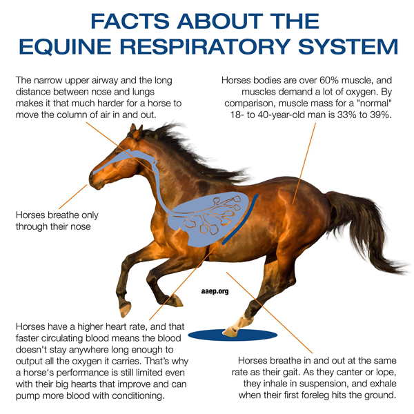
The respiratory tract begins at the nostrils. It continues through the nasal passages, pharynx, larynx, trachea, and the lungs. The nasal passages contain small bones called turbinates which are covered in respiratory epithelium that helps to remove debris and microorganisms found in the air. The paranasal sinuses are hollow spaces around the nasal passages where the reserve crown of the teeth reside. It should be noted that horses are obligate nasal breathers and cannot breathe through their mouths.
The pharynx lies beyond the nasal passages and contains the openings of the guttural pouches. These pouches hold 300-500 ml of air or purulent discharge. There are numerous blood vessels and cranial nerves in and around the pouches that can become affected from guttural pouch diseases.
Air then passes to the larynx, comprised of the epiglottis and the arytenoid cartilages. Through the arytenoids lies the rima glottidis, the potential space between the vocal ligaments contained within these intrinsic ligaments and membranes. This continues to the trachea.
The trachea is a long tube made of cartilaginous rings to conduct air to the lungs. The mucociliary apparatus of the trachea moves in a wave-like motion, helping to clear mucus and other debris from the airway, preventing lower airway infection. The trachea divides at the carina into the two mainstem bronchi, one for each lung. These subdivide into smaller and smaller bronchi and bronchioles, which lead to the air sacs or alveoli, where gas exchange takes place.
The capillaries are blood vessels that are embedded in the walls of the alveoli. Blood passes through the capillaries, brought to them by the pulmonary artery and taken away by the pulmonary vein. While in the capillaries the blood gives off carbon dioxide through the capillary wall into the alveoli and takes up oxygen from the air in the alveoli. There are approximately 10 million alveoli. The airways stretch out so that they could cover a 1,500 mile road.
The pleura are the two membranes, actually one continuous one folded on itself, that surround each lobe of the lungs and separate the lungs from the chest wall. This potential space typically contains a small amount of fluid, but when infected can contain many liters of fluid.
The horse’s respiratory tract is responsible to moving air from the atmosphere to the lungs and vice versa. The respiratory tract is divided into upper and lower portions. The upper respiratory tract is made up of the structures from the nostrils to the cervical trachea (part of trachea outside of the chest). The lower respiratory tract consists of the thoracic trachea (portion with the chest) and the lungs. Diseases of the upper and lower respiratory tract can be corrected both medically or surgically depending on the diagnosis. The key to treating any disease is to get an accurate diagnosis.
SIGNS THAT A HORSE MAY HAVE AN UPPER OR LOWER RESPIRATORY TRACT DISEASE:
- Makes noises while breathing, especially when exercising.
- Whistle
- Roaring
- Gurgling
- Rattle
- Exercise intolerant or seems to run out of energy faster than normal.
- Mucoid or bloody discharge coming from one or both nostrils
- Foul odor to his/her breathe or the horse is dropping feed.
- Large firm swelling on the horse’s face, around the eyes.
- Swelling around the throat latch region (firm or soft).
- Decreased appetite.
- Temperature greater than 101.5 F.
- Respiratory rate > 24 breaths per minute.
Any good examination begins with vital signs and routine blood work. The horse’s respiratory rate is 12-20 breaths per minute. As the respiratory tract begins at the nostrils, examination should begin there, feeling the force of air movement and noting nostril flaring. Auscultation of the trachea and lungs is next, usually employing a rebreathing bag. As we cannot ask the horse to take a deep breath, a bag is placed over the horse’s nostrils, which increases the concentration of carbon dioxide and makes the horse breathe more deeply so that the deeper regions of the lung may be heard at the surface.
Breath sounds are normally quiet. Sometimes they are loud crackles or wheezes. Sometimes they are altogether absent. Fluid sounds may be heard in the trachea.
Video endoscopy is a camera system use to evaluate the airways. The image is sent from the camera on the end of a flexible tube to a computer and monitor, allowing visualization of the respiratory structures.
This helps differentiate infectious, mechanical dysfunction, neurologic dysfunction, or traumatic disorders.
Digital radiographs are used to identify fractures or abnormalities in bones and soft tissue swelling. Frequently digital radiographs are used to identify abnormalities in the paranasal sinuses, tooth abnormalities or bone abnormalities under wounds. They are also use to evaluate the lungs for masses and abscesses.
Ultrasonography is used to diagnosis soft tissue abnormalities. It can be used to diagnosis tendon and ligament sprains, pneumonia, abnormalities in the abdomen, and identify what soft tissue structures are involved in a wound. In the lung, it is used to look for pleural fluid and solid structures, such as abscesses. Ultrasound cannot image structures deep to air in the lung, as the sound waves are not carried through air. If deeper regions of the lung can be seen, the lung is consolidated.
Diseases of the Respiratory Tract
- Idiopathic laryngeal hemisplasia (AKA paralyzed flapper)
- Arytenoid chondritis
- Dorsal Displacement of the Soft Palate
- Epiglottic Entrapment
- Subepiglottic cyst
- Paranasal sinus disease
- Primary sinusitis
- Secondary sinusitis
- Ethmoid Hematoma
- Sinus Cyst
- Neoplasia
- Pneumonia/pleuropneumonia
- Equine Asthma
Fox Run Equine Center
www.foxrunequine.com
(724) 727-3481