


UF Veterinary Hospital Offers Leading Edge Treatment to Performance Horses

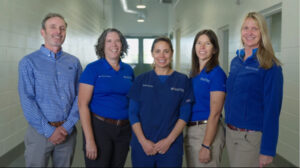
UF Veterinary Hospital at World Equestrian Center – Ocala team of board-certified specialists.
The UF Veterinary Hospital at World Equestrian Center – Ocala (WEC) offers leading-edge care to equine athletes on site as well as horses from the surrounding area, dogs and cats. No matter what brings you to WEC – Ocala, our team of board-certified specialists are here to serve you with an unprecedented array of diagnostic services.
As we enter the 2023 Winter Spectacular Show Series, we wanted to highlight some of the key services and expertise available to our equine clients. Our equine veterinarians are board-certified in equine sports medicine and are experienced performance horse veterinarians with broad skill sets, expertise and knowledge in traditional as well as novel therapies.
Well versed in the demands of each individual horse sport, our veterinarians provide a range of treatments to ensure your horse performs at its best. We also offer ACVSMR performance evaluations for lameness, purchase, neurologic, respiratory, poor performance as well as additional categories, including Soloshot videography, gastrointestinal tract examinations and telemetric electrocardiography.
A variety of rehabilitation treatments and programs are available, as well as state-of-the art diagnostic imaging. Our sports medicine specialists are supported by board-certified veterinary radiologists with specialty certification in equine diagnostic imaging who provide real-time feedback and perform complicated imaging during your horse’s visit, ensuring that we provide the highest quality imaging for your horse.
Our advanced diagnostic imaging technology includes the Qalibra CT, Hallmarq MRI, and LONGMILE PET scanner. Each tool provides different types of information and may be combined to provide more specific and accurate diagnosis of lameness and orthopedic problems.
Currently, no other facility offers the combination of these diagnostic imaging modalities at one site.
Standing computed tomography, or CT, allows us to acquire images of the horse’s head, neck, front and hind limbs without the need for general anesthesia. Our CT unit is a state-of-the art Canon Aquillion Exceed LB 160 slice scanner with a 90 cm field of view that takes just seconds to scan your horse once it is positioned. In the standing, sedated horse, the CT scanner helps diagnose lameness originating from the foot, fetlock, carpus (knee), tarsus (hock) and proximal suspensory (front and hind limbs). It is especially useful for diagnosing problems in the head such as sinusitis and dental disease as well as injuries and conditions affecting the cranial neck.
Under general anesthesia, images of the pelvis, stifle, shoulder, elbow and the most caudal portion of the neck can also be obtained.
Standing positron emission tomography, or PET, is a powerful tool that uses an injectable radioactive material, known as a radiopharmaceutical, to highlight areas of pathology that might be causing lameness from the horse’s foot to just above the carpus and tarsus. This modality is highly sensitive for detecting areas of rapid bone turnover and may provide more information than a traditional bone scan, as it acquires images in a 3-D fashion that can be fused with MRI, CT or radiographic images. The radioisotope used also has a much shorter half-life than that used in a bone scan, which means your horse has to spend less time in the hospital.
As with a bone scan, PET can be used as a screening tool if the nerve or joint blocks are not definitive about the exact region causing the lameness, your horse is not amenable to nerve or joint blocks, or if there are multiple areas of suspicion of problems on other diagnostic imaging.
We also offer standing low-field MRI, which allows us to image limbs of the horse below the carpus or tarsus while standing and sedated. MRI provides excellent soft tissue imaging, as well as information about bones and joints. This modality is useful for diagnosing any lameness condition that has been localized in the lower limb, especially in the foot, in situations where X-rays or ultrasound have not provided adequate answers. As with the CT, images from the MRI can be fused with PET scan images to provide complimentary information.
Additionally, our high-powered X-ray generator and Canon wireless DR plates allow us to obtain high quality images of regions that portable machines struggle to adequately penetrate, including the thorax (chest), abdomen, and denser areas of the spinal column.
Learn more about all of our services at hospitals.vetmed.ufl.edu/wec or contact us at (352) 414-3858 for information and appointments. Hours of operation for the equine hospital are Monday through Saturday, 8am to 5pm.