


UC Davis PET Research to Improve Horse Safety and Welfare

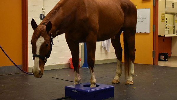
The initial standing PET prototype design enabled imaging of the hooves while the horse is under sedation, simplifying the process and facilitating the monitoring of changes over time.
A significant advance in veterinary research is coming to the racetrack in the form of a standing equine PET scanner. The machine will soon be available at Santa Anita Park, marking a major milestone in the battle against racetrack injuries.
In the wake of a difficult winter for Southern California horse racing, the entire racehorse community is looking for ways to improve safety for these amazing athletes. The approach is multifactorial and takes into consideration track conditions, medication use, training regimens and early detection of injuries. Research performed by Dr. Susan Stover, director of the J.D. Wheat Veterinary Orthopedic Research Laboratory at the UC Davis School of Veterinary Medicine, has shown that catastrophic injuries in racehorses are associated with subtle, pre-existing bone changes, particularly in the fetlock joint. The challenge has been identifying these changes early enough to prevent serious injuries.
The UC Davis imaging group has been working on this challenge of early detection of injuries for several years. What started in 2013 as a purely academic discussion between PET engineer Dr. Ramsey Badawi and equine radiologist Dr. Mathieu Spriet has led to a real-world application of the most powerful system to detect bone injuries in racehorses. The new PET scanner will be used to identify patterns of stress remodeling in the fetlock and assess how they are affected by training and track conditions. The long-term goal is to use the technique as a screening tool to identify horses at risk of catastrophic breakdowns.
“We cannot overstate how significant an advancement this is in equine diagnostic imaging, and it is a natural fit here at Santa Anita,” said Dr. Rick Arthur, equine medical director at UC Davis who advises the California Horse Racing Board on equine safety. “This is one more example of how the horse racing industry and horses benefit from working closely with UC Davis.”
Positron Emission Tomography (PET) uses a small dose of radioactivity to detect changes in bone or soft tissue at the microscopic level. Using a ring of detectors, it acquires data in three dimensions, allowing for precise detection of subtle changes, which can be early signs of compromised structures. PET can also distinguish between active and inactive lesions, which can help pinpoint areas of concern.
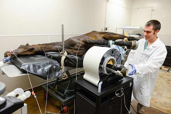
Equine PET requires the patient to be under general anesthesia since the limb needs to be still to capture the images.
The first equine PET scan was performed at UC Davis in 2015 using a scanner from Brain Biosciences, Inc. that was originally designed for human brain imaging. Horses were anesthetized, and their limbs placed in the scanner. Images were captured for up to six different areas on each horse. The results, along with support from the UC Davis Center for Equine Health and the Grayson Jockey Club Research Foundation, enabled UC Davis to initiate the first clinical equine PET program in August 2016. To date, more than 100 horses have been imaged at the UC Davis veterinary hospital for lameness evaluations.
“We are incredibly grateful to all of our donors at the UC Davis Center for Equine Health that enabled us to support research on the first equine PET unit,” said Dr. Carrie Finno, director of the Center for Equine Health. “We anticipate that this equipment will be used to prevent future injuries.”
This work demonstrated the value of PET for identifying early lesions in racehorses, but a major limitation existed: horses had to be anesthetized. Anesthesia is not an option for all patients. It requires recovery time that interrupts training, specialized equipment, and specially trained staff, as well as involves safety risks inherent in recovering horses from general anesthesia. These factors also make it difficult to perform multiple scans on a single horse in order to monitor changes over time.
With this in mind, Dr. Spriet worked with LONGMILE imaging—a division of Brain Biosciences, Inc.—to develop a scanner to image standing horses. In January 2019, the first PET scan on a standing horse was performed at UC Davis. Using an initial scanner prototype, the feet of research horses were imaged without anesthesia. This initial prototype consisted of a full ring of detectors, which would present safety issues for imaging higher parts of the limbs, such as the fetlocks. The engineers from LONGMILE designed the concept of the MILE-PET, a PET scanner consisting of a ring of detectors that open to allow quick release of the horse.
The Grayson Jockey Club Research Foundation and the Dolly Green Research Foundation showed great enthusiasm and support toward the development of the MILE-PET. Recently, the commitment of the Stronach Group to “a substantial investment in diagnostic equipment to aid in the early detection of pre-existing conditions” has made the MILE-PET scanner a reality. The new scanner will be validated this summer at UC Davis with support from the Center for Equine Health before it is installed at Santa Anita Park for the fall race meet.
“The MILE-PET is a project that has been long in the work and is now going to be deployed where it fits the best – at the racetrack at Santa Anita,” said Dr. Spriet. “It is very exciting that the product of our research, with the support of the industry, will now be able to help the fascinating world of horse racing.”