


Tendons and Ligaments of the Horse

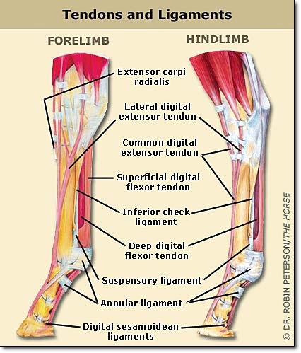
Photo provided by Fox Run Equine Center
By Brian S. Burks, DVM, Diplomate, ABVP, Board Certified in Equine Practice
Tendons and ligaments hold bones in place and help to create propulsion. Tendon and ligament injuries are common in horses performing in vigorous athletic activities. Many tendon and ligament injuries can be avoided through proper conditioning and by not pushing a horse beyond its limits. It has been estimated that about 30% of competition horses suffer from tendon damage as the result of running, jumping, and dressage.
By definition:
Tendon–A flexible but inelastic fibrous cord by which muscle is attached to bone.
Ligament–A band of tough, flexible fibrous tissue that connects bones or cartilages, serving to support and strengthen joints.
These soft tissue injuries are more common on the turf or where the footing is deep. Horses on hard surfaces tend to damage joints and bones, rather than tendons or ligaments, though these may be secondary to catastrophic bone injury.
Jumping stretches tendons to the limit and beyond when a horse lands following a six-foot jump. The fetlock touches the ground under such circumstances.
The deep digital and superficial flexor tendons play an important role in the horse’s movement. The suspensory ligament, which originates at the upper end of the third metacarpal bone and the lower edges of the distal row of carpal bones is also quite important.
Tendons and ligaments have a similar structure, but tendons tend to have more stretch capability compared to ligaments. Both are made from collagen fibers arranged in linear fashion, which allows stretching.
Collagen is a tough protein found in skin, tendons, bone, cartilage, ligaments and other connective tissues. It allows tendons and ligaments to stretch and contract as well as provide their sturdiness. The collagen fibers are put together much like ropes and are cross-linked to provide additional stability.
The superficial flexor tendon is readily visible as it runs down the back of the cannon bone close to the skin. It originates at the superficial flexor muscle just behind the elbow on the medial humeral epicondyle in the front legs and from the proximal tibia near the stifle in the rear limbs. It inserts on the middle phalanx, where it flexes the proximal and middle phalangeal joints, and stabilizes metacarpophalangeal (fetlock) joint.
The deep digital flexor tendon arises as three muscle bellies from its origin on the medial humeral epicondyle, fusing to form a common tendon just above the carpus on the caudal aspect of the limb. The single tendon passes distally, enclosed in the carpal sheath, through the carpal canal. In the mid-metacarpal region, the tendon is enforced by the distal accessory ligament (inferior check ligament) which limits the movement of the tendon, preventing over stretching. At the metacarpophalangeal (fetlock) joint, the DDFT passes over the sesamoid groove. In the middle of the proximal phalanx, or long pastern bone, the DDFT runs between the branches of the SDFT and over the distal sesamoid (navicular) bone to insert on the distal phalanx.
Flexor tendons help the leg and foot move in a rearward motion, whereas the extensor tendons move the limb forward. More stress is placed on the flexor tendons. As the limb bears weight the carpus overextends, stretching the flexor tendons, so that the two flexor tendons are more commonly injured with strain injuries or tendinitis- a bowed tendon is tendinitis of the SDFT, where there is tearing, inflammation, and swelling cause the tendon to bulge, or bow, outward.
The suspensory ligament, or interosseous muscle, is a strong, flat ligament with variable amounts of muscle fibers. It originates from the back of the carpus (“knee”) and extends the length of the canon bone, between the splint bones. Below the fetlock, it becomes several other ligaments, known as the distal sesamoidean ligaments, which insert on the pastern. The SL acts as a spring, storing and releasing energy during movement. It prevents hyperextension of the fetlock. If this ligament becomes torn, the fetlock will drop toward the ground during normal weight bearing.
Each tendon is enclosed in a sheath wherever there is a change in direction or there is likely to be friction during movement. There is a carpal sheath, fetlock sheath and a pastern sheath. The sheath has synovial fluid to help eliminate friction as the tendon moves. Tendons also serve as shock absorbers, dissipating concussive forces that would otherwise be given to joints, bones, and muscles. Stress, or load, occurs during weight bearing of an individual limb, which is accommodated by lengthening of the tendon.
During exercise, the tendons of a horse can stretch from one to three inches. When the tendon is stretched beyond its strain capacity, the collagen fibers tear, resulting in inflammation, pain, and loss of normal function.
Most serious injuries occur on the forelimbs as 60% of the body weight is borne by the front end. There is also a point where a single forelimb bears the entire weight of the horse, putting a great deal of strain on the tendons and suspensory apparatus. When jumping, all the concussion is taken by the front limbs.
Prevention of tendon injuries
The top three preventive steps are: 1) conditioning, 2) conditioning and 3) conditioning! The horses at the highest risk of injury are those that are unfit or have had a period of lay-off and are then suddenly worked hard. It is important to work with a good trainer or educate yourself on the best way to condition a horse for its intended use.
Build layers of fitness gradually and consistently. Never work to the point of exhaustion/fatigue, as this is when tendon-ligament injury is most likely to occur. Only after the horse is fit should you gradually add discipline-specific rigors, such as collection in a dressage horse, speed and tight turns in a barrel horse or uneven terrain in a trail horse.
Other preventive efforts include regularly palpating your horse’s legs before and after exercise to monitor for heat and swelling; regular farrier care to maintain good foot balance; warming up before and cooling down after strenuous exercise; and good footing during hard work whenever possible. Stop exercise immediately if your horse feels “off,” and call an equine veterinarian familiar with sports medicine sooner rather than later.
Start out slowly and build gradually. This also helps warm up your muscles. Maintain your horse at a healthy weight; overweight horses put more stress on their joints, tendons, and ligaments.
Vary your routine with a balance of cardio exercise and strength training. After intense exercise, give your horse a day off, or at least switch to a different activity. That helps reduce the risk for over-stressing the same ligaments and tendons.
Stretch. Stretching after exercise, when the body is warm and more pliable helps maintain a tendon’s ability to elongate during exercise. Hold a stretch for no more than 10 to 20 seconds.