


Study Links Bone Loss to Proximal Sesamoid Bone Fractures

UC Davis School of Vet Medicine
by: Amy Young
A recent study by Sarah Shaffer, Dr. Susan Stover and colleagues at the J.D. Wheat Veterinary Orthopedic Research Laboratory at the UC Davis School of Veterinary Medicine sought to characterize bone abnormalities that precede proximal sesamoid bone (PSB) fractures and determine if pre-existing abnormalities are associated with these fractures. The group retrospectively studied cases from California Thoroughbred racehorses that died from PSB fractures, and controls that died for other reasons.
The most common fatal injury in racehorses in the United States, PSB fractures account for 45-50 percent of such injuries in Thoroughbreds, and 37-40 percent in racing Quarter Horses. The PSBs are two comparatively small bones located in the fetlock that act as part of the suspensory apparatus. Fractures in these bones are likely due to the accumulation of repeated, stress-related processes. This is supported by evidence that racehorses in intensive training are at higher risk for PSB fractures, but the exact causes are not well understood.
Other repetitive overuse injuries in horses are known to be bilateral in nature, meaning that they are similar on both sides of the horse, with the more severely affected limb usually incurring the fracture. With this in mind, the study looked at both the fractured PSB and the intact PSB from the opposing limb of the same horse for all of the cases. The researchers hypothesized that horses with PSB fractures would also show evidence of stress in the PSB of the opposite limb and that the bone that sustained the break would show more severe changes than the intact bone.
The results showed that 90 percent of fractured PSBs from the cases had visible discoloration on the surface of the fracture, most commonly (70 percent of the time) in a characteristic crescent pattern. Directly below the cartilage, evidence of bone loss was noted in 70 percent of cases. This bone loss was located in the same region as the discolorations. Fractured PSBs had lower bone volume fraction and tissue mineral density within the lesion sites than comparable locations in opposing limbs and controls. These regions were contiguous with the fracture lines. Evidence of microdamage was also observed in fractured PSBs.
Overall, changes identified in the bones were more numerous in case horses than control horses and more severe in the fractured limbs than the opposing limbs in cases. Sampling from areas of bone distant from the lesions noted no significant differences in bones from case and control horses other than the presence of a lesion.
This data supports the role of microdamage and tissue remodeling in the formation of lesions in PSBs. It is important to note that all of the horses in this study were California racehorses, so it is currently unknown if the results will apply equally to racehorses in other areas. Future studies with larger sample sizes may provide further information.
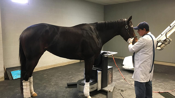
Understanding the mechanism of PSB fracture is necessary in order to determine risk factors and prevent fractures. Combining this information with advanced technology, such as the recent introduction of positron emission tomography (PET scan) may facilitate identification of horses at risk for PSB fracture and inform management alterations to avoid injury.