


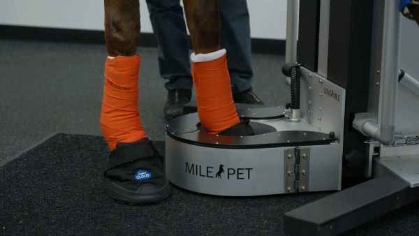
Here at the UF Veterinary Hospital at WEC, we are the only facility in the area to offer PET scan along with standing CT and standing MRI available in the same hospital.
The PET machine itself is similar to a traditional bonescan machine, in that it uses an injectable radioactive substance (radioisotope) to show up regions of potential pathology in either bone or soft tissues. It has a number of advantages over bonescan, one of the biggest being the ability to ‘fuse’ the PET images with both MRI and CT images to precisely identify the abnormalities. The standing PET scanner also allows us to acquire images in 3D rather than just in two dimensions, which is one of the major shortcomings of bone scan. Additionally, as the radioisotopes have a much shorter half-life than the those used in bonescan, horses can be discharged from the hospital on the same day, eliminating the need for an overnight hospital stay after scanning.
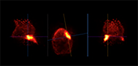
Photo by UF Veterinary Hospital.
On its own, the PET scanner can help to identify active areas of pathology that might be causing lameness. It is especially useful when nerve blocks have isolated the region where the lameness might be coming from, but there is possibly more than one site of pathology in that area. It is also very useful in helping us decide which imaging modality is better to make a more definitive diagnosis, such as standing MRI or CT.
When the PET is combined with either standing MRI or standing CT, it exponentially increases the chances of making a definitive diagnosis by allowing us to know what is happening metabolically in this area. This information can be invaluable in helping us to provide prompt and accurate therapy, and getting your horse back in the ring!
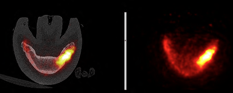
Photo by UF Veterinary Hospital.
This coming year there will be a clinical study, in which horses performing in dressage, jumping or three-day eventing will be eligible. To be enrolled, horses will need to have lameness that has been localized to the fetlock region based on regional analgesia. Horses must be scheduled to have a standing MRI of the fetlock and have had improvement of the lameness following a block of the fetlock. Also eligible are horses who present for standing fetlock MRI, whose lameness did not improve with a palmar digital nerve block, but did improve after abaxial sesamoid nerve block, but there was no pathology identified on foot or pastern MRI to explain the clinical signs.
Learn more about all of our services at hospitals.vetmed.ufl.edu/wec or contact us at 352.414.3858 for information and appointments. On Facebook? Follow UF Veterinary Hospital at WEC.