


Legs- Common Injuries and How We Treat Them

By: Dr. Brianne Henderson
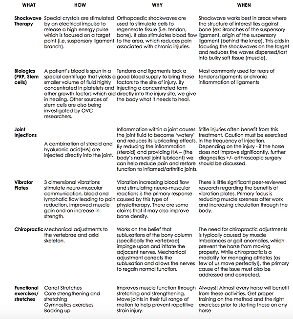
Every year, riders are plagued with limb injuries to their equine partners. Sometimes, this is a small blip in the training schedule. Other times, it spells the end of a competitive season. In this article we are going to highlight some common injuries and different modalities that can help your horse onto the road to recovery (and a few preventative tips!). At the end of the article, there is a table outlining different treatments to help manage the injuries discussed.
The Suspensory Ligament
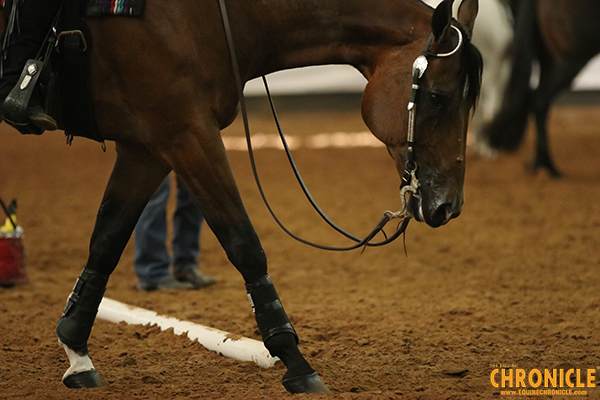
The Suspensory Ligament (SL) is effectively a broad elastic cable that runs from the back of the knee down the back of the canon bone and then branches to cradle the fetlock joint. Its purpose is to suspend the fetlock joint and prevent over extension during loading. Essentially, it works like a spring mechanism to absorb the energy as it stretches and then recoils to lift the fetlock back to its normal position.
Over time, micro-damage occurs within the fibres of the ligament and due to the poor blood supply, these micro-tears are more likely to accumulate than heal. Often this is why we see repetitive strain injuries/inflammation to the SL. The impact of foot balance is critical to the health of the SL. A foot with long toe/low heel places increased strain on structures within the foot (navicular bone), the Deep Digital Flexor Tendon and the Suspensory ligament by predisposing to a “toe first” landing. Instead of allowing a gradual absorption of impact through the structures of the heels and up the limb, this movement creates a snapping action on the DDFT and Suspensory ligament. The long-term effect of foot imbalance is associated with chronic heel pain (“toe first”) and also chronic proximal suspensory desmitis.
Veterinary treatment options may include shockwave therapy and biological injections.
Stifle
The stifle is both the largest and the most complex synovial joint of the horse and due to its location and structure, it is also the most vulnerable. In comparison, the stifle is most similar to the human knee.
The stifle consists of two joints:
Femoropatellar joint is formed between the femur and the joint surfaces of the patella. It has multiple ligaments that aid in the support and function of this joint.
Femorotibial joint is formed by the back surface of the femur (condyles) and the tibia.
The menisci are small wedge shaped “cushions” that lie between the femur and the tibia and provide a shock absorbing function.
Collateral ligaments – lie outside the stifle joints and connect the femur to tibia and femur to patella.
Cruciate Ligaments – lie within the femorotibial joint and stabilize the tibia against the femur
The most common injuries to the stifle in sport horses are related to the soft tissue structures (ligament injuries, cruciate ligament tear and meniscus damage) and secondary arthritis within the joint. Often, these injuries are the direct result of trauma that results in multiple injuries to one joint. For example a Combined Training horse that crashes or has a rotational fall would be at risk of not only a medial meniscus tear but also cartilage damage and collateral (supportive) ligament damage. Return to full function depends on the severity of damage but can range from 10% to 50% (Dyson). Early diagnosis through joint blocks, x-ray and ultrasound will enable a treatment and rehabilitation program to be put in place as soon as possible. Horses that do not respond to medical management (stem cell injections, steroid injections) may require surgery to fully understand the extent of the injury as well as reduce the debris floating inside the joint.
Veterinary treatment options may include biological injections (stems cells), joint injections, and arthroscopic surgery.
Sacro-Iliac Damage
Dressage and show jumping horses are more commonly affected by Sacro-Iliac (SI) pain/inflammation. Common complaints from riders range from a sudden unwillingness to go forward when ridden to bucking/kicking when ridden. Other riders will grow concerned over reduced impulsion and engagement (worse at canter than trot) or a “stiff backed” horse. With true SI pain the signs are almost always worse when the horse is being ridden as they are focused on reduced power and suppleness.
Anyone who has suffered from lower back pain understands how debilitating it can be, but what does SI pain mean? Typically, SI pain is secondary to another lameness issue in the horse or a result of repetitive strain. Common primary issues would be hind limb lameness and back pain under the saddle (poor saddle fit, kissing spines). These injuries prevent the horse from using their bodies normally and places abnormal stress on the SI joint eventually causing pain.
Proper identification and treatment of primary lameness issues must be the first step in getting a horse on the road to recovery; however, functional exercises play a major role in maintaining the health of the SI joint. The use of carrot stretches, gymnastics, walking over poles and walking up and down hills all move the SI joints in their full range of motion and stretch the joint and soft tissue associated with the joint. It is restricted range of motion that causes pain/stiffness and eventually abnormalities/injury of the structures within and around the joint.
Treatment options may include: carrot stretches, functional exercises, and joint injections. Discuss possible chiropractic treatment with your vet.
When faced with a major injury, there are many modern treatments and therapy modalities that can help return your horse to work. However, we must remember that one of the most important factors when healing an injury is “time.” If a horse is pushed back into work without appropriate rest time, they will be at risk of injuring themselves again. Working alongside your veterinarian to ensure an accurate diagnosis and appropriate treatment program is essential to getting your horse on the right road to recovery!
Many of the injuries encountered in athletic horses are the result of repetitive strain/overuse or speed. This highlights the need for cross training and functional exercises/stretches to be a regular part of a horses’ training in order to reduce the risk of this type of injury. Riders are encouraged to implement carrot stretches and functional exercises into their daily/weekly routine!
Sign up for our free e-newsletter which will deliver monthly welfare tips and announce tools to aid all horse owners in carrying out their ‘Full-Circle-Responsibility’ to our beloved horses. In partnership with the Ontario Ministry of Agriculture and Food, Equine Guelph is developing a ‘Full-Circle-Responsibility’ equine welfare educational initiative which stands to benefit the welfare of horses in both the racing and non- racing sectors.
Visit Equine Guelph’s Welfare Education page for more information.
See the other articles in this series:
Part 1 – Developing the Sport Horse: The importance of cross training for mind and body
Part 2 – Developing the Sport Horse: the importance of hydration