


At the Forefront of PET Veterinary Imaging

 UC Davis School of Veterinary Medicine
By: Rob Warren
Positron emission tomography (PET) scanning technology has now been in place at the UC Davis veterinary hospital for two years. In 2016, UC Davis became the first veterinary hospital in the world to implement an equine PET scanner, and has since added a small animal scanner in 2018.
The program has enjoyed a successful second year with many exciting developments.
One of this year’s most important achievements was the validation of a dual-tracer scanning protocol, which allows for the assessment of both bone and soft tissue lesions in lame horses. This protocol has now been used on several clinical cases and has been utilized to assess complex lesions involving the foot and the suspensory ligament. (Figure 1)
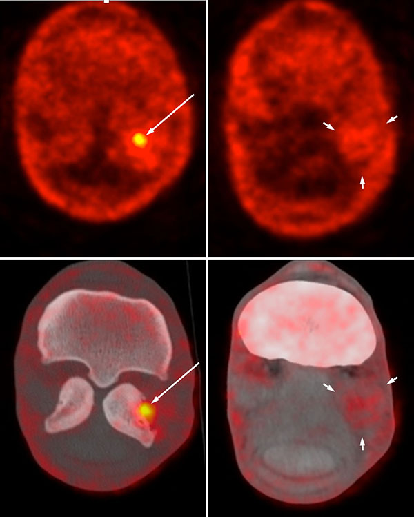
Figure 1: NaF PET and fused PET CT transverse images of the fetlock of a 6-year-old Warmblood horse. There is marked focal increased radiotracer uptake in the lateral proximal sesamoid bone (long arrow) and diffuse moderate increased uptake through the lateral branch of the suspensory ligament (short arrows). This demonstrates an active desmitis of the lateral branch with focal insertional desmopathy.
PET has also proven to be an interesting component for laminitis research. Laminitis, as most horse owners know, is an extremely serious condition of the horse foot that, unfortunately, is often fatal. There are still a lot of unknowns on the origin and development of the disease. While researchers continue to investigate the disease, PET has brought new insights on the areas of the foot that are the most affected. (Figure 2)
“With PET tracking, we are now able to confirm the presence of stem cells in a specific lesion in a tendon,” said Dr. Spriet. “Using scintigraphy, we were limited to only being able to tell that stem cells were present in the region of the foot.”
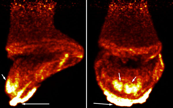
Figure 2: 3D maximal intensity projection of the foot of a 15-year-old American Quarter Horse mare with severe laminitis. There is moderate increased uptake at the dorsal distal aspect of the hoof wall (short arrow) and marked FDG uptake through the sole (long arrow). This indicates increased metabolic activity in the lamina at the site of rotation and marked inflammation as the toe of the distal phalanx protruded through the sole.
In addition to multitude of research studies, UC Davis has scanned more than 40 clinical patients, bringing useful information for management of cases. In one case, Irish Streetsinger—a racehorse with persistent lameness—was admitted to UC Davis. Following several lameness evaluations (including radiographs and nuclear scintigraphy to determine the area of lameness), clinicians advised her owner that a PET scan would be the best option to obtain a conclusive diagnosis. The scan pinpointed her lameness, and following stem cell treatment, she was back on the track. In 2018, Irish Streetsinger ran seven races with one victory.
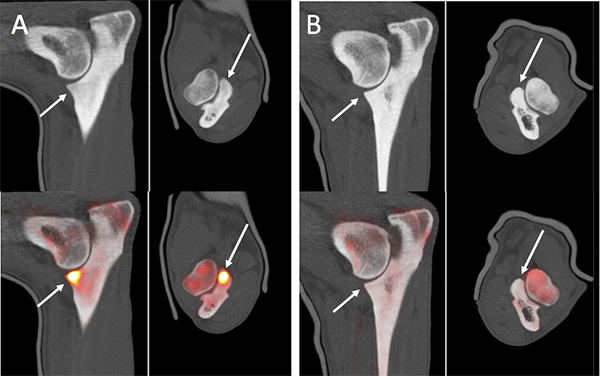
Figure 3: Multiplanar fused NaF PET and CT images of a 1-year-old Labrador dog with pain in the right elbow. The CT images showed mild bilateral remodeling of the medial coronoid process. The PET images demonstrated marked 18F-NaF uptake in the right medial coronoid process (A). The left medial coronoid process (B) did not have any abnormal 18F-NaF uptake. This correlated well with the clinical signs of right elbow pain.
Drs. Spriet and Pablo Espinosa have presented their PET research at multiple regional, national and international conferences, including annual meetings of the American College of Veterinary Radiology, the European College of Veterinary Diagnostic Imaging, and the Society of Nuclear Medicine and Molecular Imaging.
“We are seeing a tremendous amount of enthusiasm for the use of PET from the veterinary imaging community,” said Dr. Spriet. “Members of veterinary imaging organizations from around the world are excited to learn about our advancements for the capabilities of equine PET imaging.”
This second year of the PET program at UC Davis has also seen the development of a canine PET program. Using the piPET scanner, two research projects were completed: one assessing the regional extension of tumors of the oral cavity and one evaluating elbow pain.
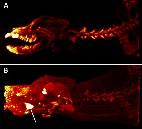
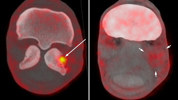
Figure 4: Maximal intensity projection NaF images (A) and FDG images (B) of a 5-year-old Labrador dog with chronic pain on opening the mouth. There is no significant osseous lesion except for moderate periodontal disease. The FDG images demonstrated marked focal increased uptake in the tongue that was secondary to accumulation of “fox-tails” in the tongue and abscessation (arrow).
PET is most commonly used in human medicine to assess the extent of tumors and the presence of metastasis. A clinical trial at UC Davis for the treatment of oral tumors in dogs confirmed the value of PET scanning for treatment planning.
A final highlight of the year was the installation of the Mini Explorer II small animal PET scanner, making UC Davis the only veterinary hospital in the world with a volumetric PET scanner. This project was made possible through the vision of Dr. Erik Wisner, associate director of the UC Davis veterinary hospital in charge of diagnostic imaging services, and grant support from the UC Davis Center for Companion Animal Health.
The installation came about through a collaboration with Drs. Ramsey Badawi and Simon Cherry of the UC Davis Medical Center and the Department of Biomedical Engineering, respectively, who have designed this specific scanner as a prototype for their NIH-funded whole body PET scanner, the Explorer.
“Through our robust clinical and research programs over the past two years, UC Davis has positioned itself at the forefront of veterinary PET imaging,” said Dr. Wisner. “We are leading the way in the development of novel imaging techniques and providing our clients cutting-edge clinical care.”