


Advancements in Equine Imaging
Click here to read the complete article
328 – October, 2016
BY MEGAN ARSZMAN
 The world of medicine is always evolving. It doesn’t matter if it’s human, canine, or equine—new ideas and concepts are being tried and tested on a daily basis, all with the goal of improving health. Sometimes, developments have a trickle-down effect, helping other species wherein there was no initial intention. This has been the case with recent advancements in equine imaging: CT scanners, positron emission tomography scanners, threedimensional imaging, and 360-degree digital radiographic studies. University of California, Davis
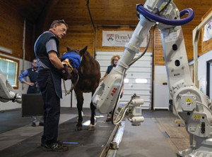
The ability to put a timeline on an injury provides helpful information, not only for diagnosing but also treatment, thanks to the new positron emission tomography (PET) scanner that’s available at the University of California, Davis School of Veterinary Medicine’s Center for Equine Health. The teaching facility is the first veterinary facility in the world to utilize this imaging technology for equine patients, and researchers are just starting to discover the machine’s capabilities.
Mathieu Spriet, DVM, MS, Associate Professor in Surgical and Radiological sciences at UC Davis, has been interested in using the PET scanner on horses for a few years. What really sparked his interest was when he read a paper about how researchers were using a PET/CT scanner in comparison with an MRI to look into obscure foot pain in people. “It struck me because obscure pain is what we deal with every day,” he says.
The main difference between a PET scanner and other imaging modalities is that a PET scanner is a “functional” imaging technique—meaning it can detect changes in the tissue because it observes activity at the molecular level. Other imaging techniques discover “morphological” information, because they can only identify changes in the size, shape, or density of structures. With a PET scanner, not only can you detect lesions missed with other modalities, but you can also tell if an injury is “active” (meaning just forming), rather than an old injury that may have been there for months or even years and shows up on other modalities as scar tissue.
“If you see something on a PET scanner, you know it’s an injury that’s active now,” Dr. Spriet says. “It helps to understand the meaning of different lesions, and it’s a great tool to use to follow the evolution of the lesion.”
Click here to read the complete article328 – October, 2016